Improved graft survival by using three-dimensional printing of intra-abdominal cavity to prevent large-for-size syndrome in liv
Author
jsrrules
Date
2024-12-08 18:53
Views
1123

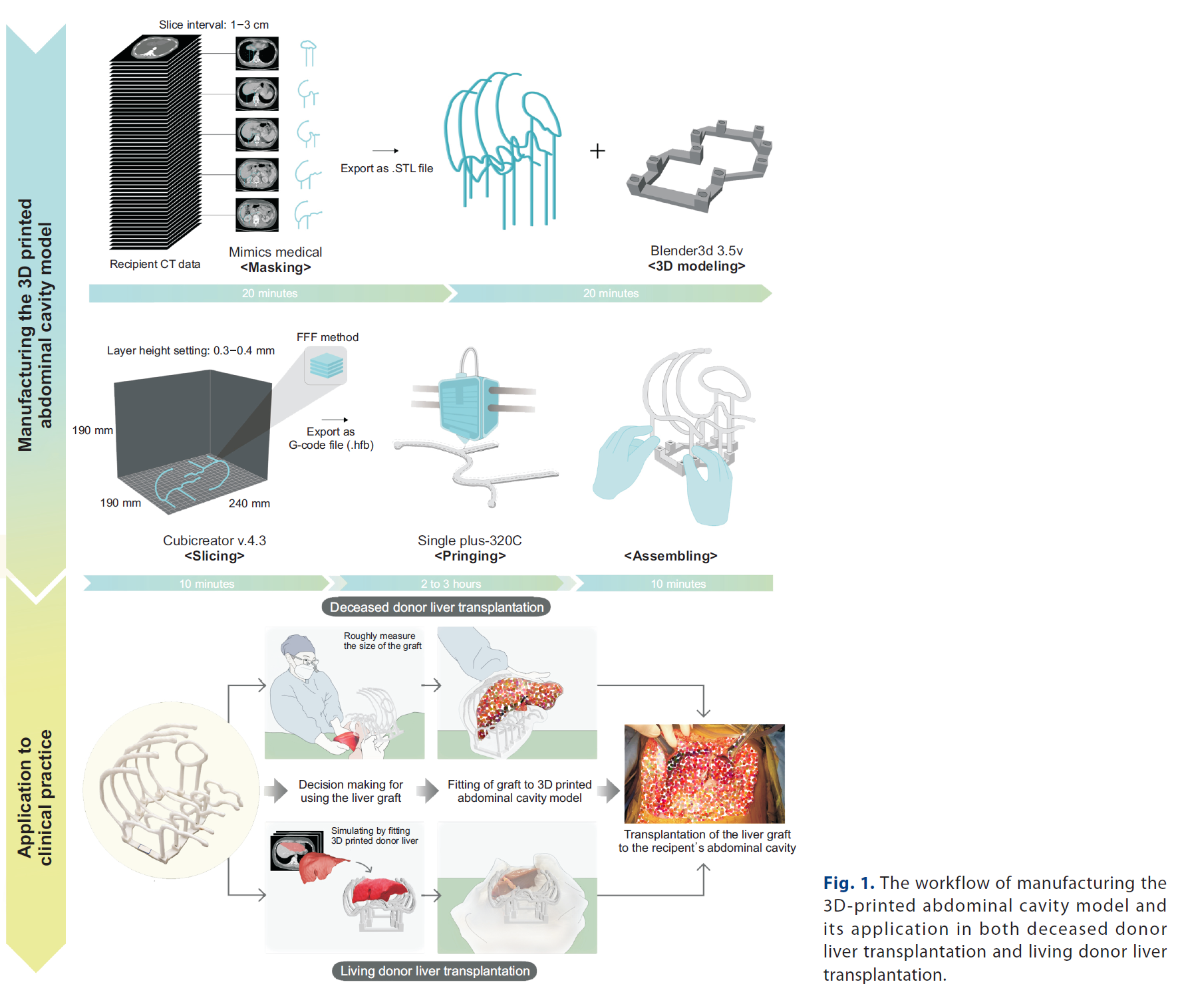

Backgrounds/Aims: While large-for-size syndrome is uncommon in liver transplantation (LT), it can result in fatal outcome. To prevent such fatality, we manufactured 3D-printed intra-abdominal cavity replicas to provide intuitive understanding of the sizes of the graft and the patient’s abdomen in patients with small body size between July 2020 and February 2022.
Methods: Clinical outcomes were compared between patients using our 3D model during LT, and patients who underwent LT without 3D model by using 1 : 5 ratio propensity score-matched analysis.
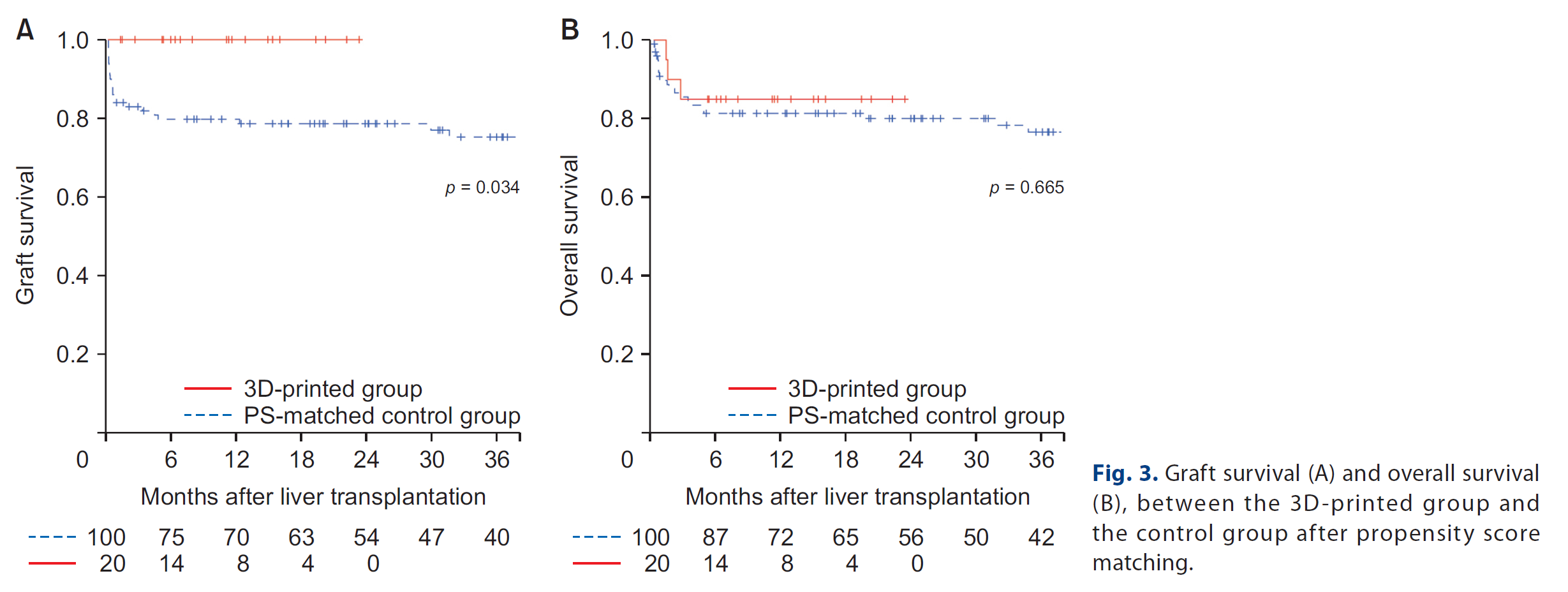
Results: After matching, a total of 20 patients using 3D-printed abdominal cavity model and 100 patients of the control group were included in this study. There were no significant differences in 30-day postoperative complication (50.0% vs. 64.0%, p = 0.356) and the incidence of large-for-size syndrome (0% vs. 7%, p = 0.599). Overall survival of the 3D-printed group was similar to that of the control group (p = 0.665), but graft survival was significantly superior in the 3D-printed group, compared to the control group (p = 0.034).
Conclusions: Since it showed better graft survival, as well as low cost and short production time, our 3D-printing protocol can be a feasible option for patients with small abdominal cavity to prevent large-for-size syndrome after LT.
This study explores the use of 3D-printed abdominal cavity models in liver transplantation (LT) to address large-for-size syndrome, which can lead to fatal outcomes. A total of 20 patients underwent LT using 3D-printed models, and their outcomes were compared with a control group of 100 patients via propensity score matching.
Key Findings:
- Graft Survival: The 3D-printed group had significantly superior graft survival compared to the control group.
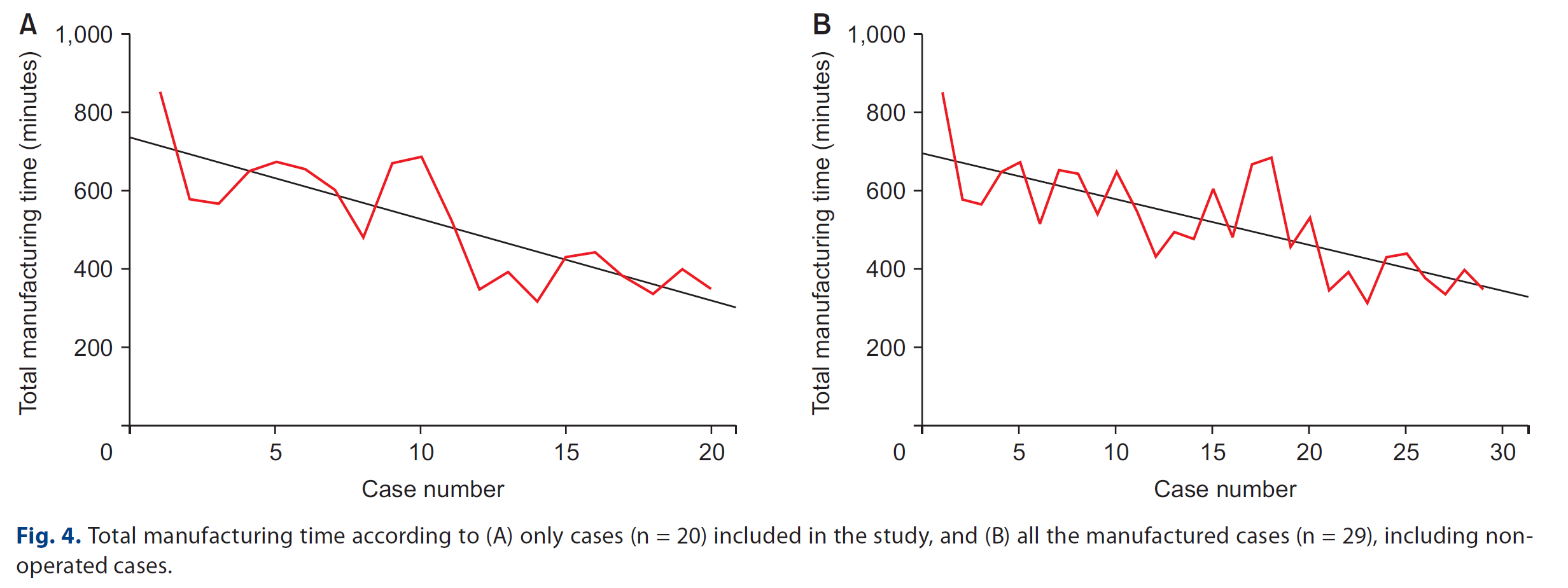
- Cost and Time Efficiency: Manufacturing cost was approximately $1.25 per model, with a production time of about 6-7 hours, showcasing cost-effectiveness and practicality.
- Applications: The models were particularly helpful for recipients with small abdominal cavities, aiding in selecting appropriate grafts and preventing complications.
Conclusion: The integration of 3D-printed models into clinical practice improves surgical planning and outcomes while being cost-effective and efficient.
3D printing technology is redefining surgical approaches, providing personalized anatomical models for preoperative planning and intraoperative guidance. Its application in liver transplantation exemplifies its potential to improve graft selection, prevent complications, and enhance surgical outcomes. With advancements in cost and production time, 3D printing is becoming an accessible tool for precision medicine.
이 연구는 간 이식(LT) 중 과대 크기 증후군을 예방하기 위해 3D 프린팅 복강 모델을 활용한 사례를 다룹니다. 3D 프린팅 모델을 이용하여 간 이식을 받은 20명의 환자와 100명의 대조군 환자 결과를 성향 점수 매칭을 통해 비교했습니다.
주요 결과:
- 이식 생존율: 3D 프린팅 모델을 사용한 그룹이 대조군에 비해 이식 생존율이 유의미하게 높았습니다.
- 비용 및 시간 효율성: 모델 제작 비용은 약 $1.25, 제작 시간은 약 6~7시간으로 비용 효율성과 실용성을 보여주었습니다.
- 적용 사례: 특히 작은 복강을 가진 수혜자들에게 적합한 간을 선택하고 합병증을 예방하는 데 유용했습니다.
- 결론: 3D 프린팅 모델의 임상 적용은 수술 계획과 결과를 개선하며 비용 효율적이고 실용적인 방법임을 입증했습니다.
3D 프린팅 기술은 수술 접근 방식을 혁신하며, 수술 전 계획 및 수술 중 가이드를 위한 개인 맞춤형 해부학적 모델을 제공합니다. 간 이식 분야에서의 활용은 이식 간 선택을 개선하고 합병증을 예방하며 수술 결과를 향상시키는 잠재력을 보여줍니다. 비용 및 제작 시간의 발전으로 3D 프린팅은 정밀 의학의 도구로 점점 더 접근 가능해지고 있습니다.
📥 다운로드 횟수: 50회