Automated 3D liver segmentation from hepatobiliary phase MRI for enhanced preoperative planning
Author
jsrrules
Date
2024-12-13 15:28
Views
389

Recent advancements in deep learning have facilitated significant progress in medical image analysis. However, there is lack of studies specifically addressing the needs of surgeons in terms of practicality and precision for surgical planning. Accurate understanding of anatomical structures, such as the liver and its intrahepatic structures, is crucial for preoperative planning from a surgeon’s standpoint. This study proposes a deep learning model for automatic segmentation of liver parenchyma, vascular
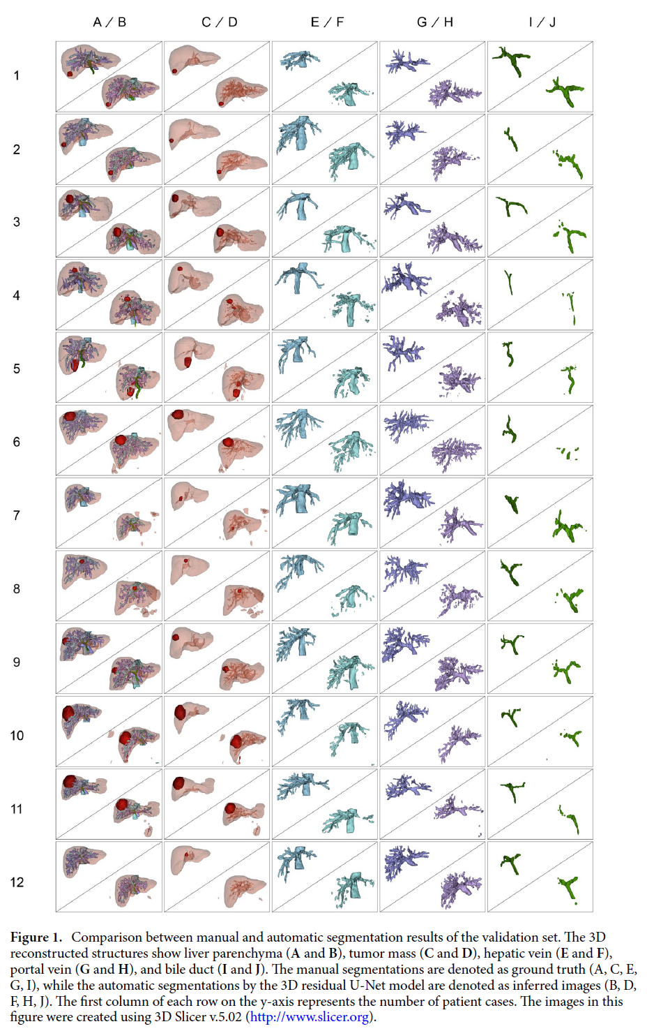
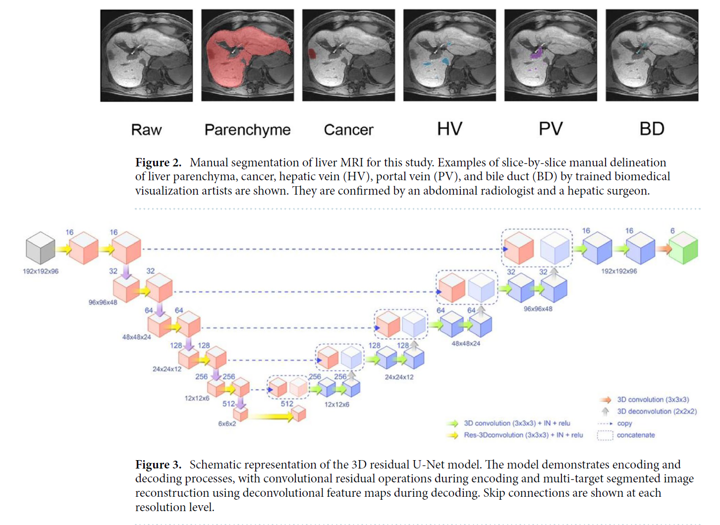
and biliary structures, and tumor mass in hepatobiliary phase liver MRI to improve preoperative planning and enhance patient outcomes. A total of 120 adult patients who underwent liver resection due to hepatic mass and had preoperative gadoxetic acid-enhanced MRI were included in the study. A 3D residual U-Net model was developed for automatic segmentation of liver parenchyma, tumor mass, hepatic vein (HV), portal vein (PV), and bile duct (BD). The model’s performance was assessed
using Dice similarity coefficient (DSC) by comparing the results with manually delineated structures. The model achieved high accuracy in segmenting liver parenchyma (DSC 0.92 ± 0.03), tumor mass (DSC 0.77 ± 0.21), hepatic vein (DSC 0.70 ± 0.05), portal vein (DSC 0.61 ± 0.03), and bile duct (DSC 0.58 ± 0.15). The study demonstrated the potential of the 3D residual U-Net model to provide a comprehensive understanding of liver anatomy and tumors for preoperative planning, potentially leading to improved surgical outcomes and increased patient safety.
심층 학습의 최근 발전은 의료 영상 분석에서 상당한 진전을 가능하게 했습니다. 그러나 수술 계획의 실용성과 정확성 측면에서 외과의의 요구를 구체적으로 다룬 연구는 부족한 실정입니다.
외과의의 관점에서 간 및 간내 구조와 같은 해부학적 구조를 정확히 이해하는 것은 수술 전 계획에 매우 중요합니다. 본 연구는 간담췌 단계 간 MRI에서 간 실질, 혈관 및 담도 구조, 종양 덩어리를 자동으로 분할하기 위한 심층 학습 모델을 제안하여 수술 전 계획을 개선하고 환자 결과를 향상시키는 것을 목표로 합니다.
총 120명의 성인을 대상으로 연구를 진행했으며, 이들은 간 종양으로 인해 간 절제술을 받았고, 수술 전 가독세트산 강화 MRI를 시행하였습니다. 간 실질, 종양, 간 정맥(HV), 문맥(PV), 담도(BD)를 자동으로 분할하기 위해 3D 잔여 U-Net(Residual U-Net) 모델을 개발하였고, 수작업으로 표시된 구조와 비교하여 Dice 유사도 계수(DSC)를 사용해 모델의 성능을 평가하였습니다.
모델은 간 실질(DSC 0.92 ± 0.03), 종양 (DSC 0.77 ± 0.21), 간 정맥(DSC 0.70 ± 0.05), 문맥(DSC 0.61 ± 0.03), 담도(DSC 0.58 ± 0.15)의 분할에서 높은 정확도를 달성했습니다.
본 연구는 3D 잔여 U-Net 모델이 간 해부학 및 종양에 대한 포괄적인 이해를 제공하여 수술 전 계획을 개선하고, 궁극적으로 수술 결과를 향상시키며 환자 안전성을 높이는 데 기여할 가능성을 입증하였습니다.
📥 다운로드 횟수: 50회